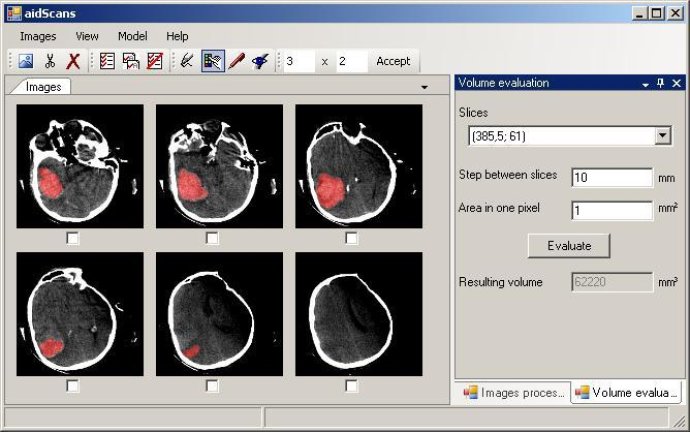
This software is designed for volume estimation and medical imaging, specifically for calculating brain tumor volumes using the input of scanned object slices. It supports open interfaces for input and output formats.

One of the standout features of this application is that it supports open input/output format interfaces. This allows for easy and fast integration with custom file formats and data exchange with existing applications. Therefore, users can seamlessly incorporate it into their workflows without having to worry about compatibility issues.
The application's key features include a multi-image working area that enables manipulation and editing of similar slices. Additionally, the program offers window/center correction, known as brightness/contrast adjustment, as well as a manual selection tool. Users can also take advantage of the auto-selection tool, which helps identify solid sub-parts with similar density such as tumors, hollow spaces, organs, and more. A border tool is also included for manual separation of different sub-parts with similar density.
Other features of the program include a volume and area estimation dialog, a 3D-model view of the selected volume, and input/output formats openness which allows easy integration with existing software while supporting popular and custom data formats.
Overall, the volume estimation application is a robust program that offers a wide range of features for users. With its versatility, ease of use, and compatibility with various data formats, this software is a valuable tool for medical imaging and other industries like geology.
Version 4.7.1: Distances estimation and slice area chart were added
Version 4.8: Distances estimation and slice area chart were added
Version 4.6: Support of most popular DICOM files and animation of angiograms are added
Version 4.5: Export to AutoCAD DXF-files was added
Version 4.4: 3D-model view